MOHS MICROGRAPHIC SURGERY
 What Is Mohs Micrographic Surgery?
Mohs micrographic surgery (also known as Mohs chemosurgery) is a state-of-the-art treatment for skin cancer in which the diseased area is removed with the accuracy and precision of a microscope. Of all techniques to remove skin cancer, Mohs micrographic surgery has the highest cure rate and minimizes the extent of scarring. Mohs surgery is a one day, outpatient treatment which is generally reserved for use only with certain skin cancers. Those that are large, have recurred following conventional treatment, or those found in certain anatomic locations such as the central face are very effectively treated with this technique. Mohs surgery is utilized most often for the treatment of basal cell carcinoma, squamous cell carcinoma, and lentigo maligna (an early form of melanoma usually occurring on the face). However, the Mohs technique is also useful for other, less common forms of skin cancer, including sebaceous carcinoma, merkel cell carcinoma, atypical fibroxanthoma, microcystic adnexal carcinoma and dermatofibrosarcoma protuberans.
In the Mohs micrographic method, the doctor serves as surgeon, pathologist, and reconstructive surgeon. The technique, created by Dr. Frederick Mohs, is as follows:
- A local anesthetic (usually lidocaine) is injected around the skin cancer.
- The visible portion of the skin cancer is surgically removed.
- The physician then removes a thin layer of tissue around the edges and at the base of the cancer.
- The tissue is dyed to distinguish between top and bottom and left from right. A drawing, or map, is made so that the physician can pinpoint any areas of persistent tumor.
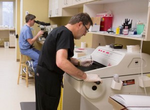
- The tissue is then sectioned into very thin slices so that the edges and underside can be examined with a microscope.
- If the tissue is found to still contain cancer, the physician outlines on the map the exact areas of involvement. Additional tissue is then removed only from the specific area(s) of involvement, thereby preserving as much healthy tissue as possible and minimizing the degree of scarring. The specimen is again processed in the laboratory, and the cycle is repeated as many times as necessary until no cancer cells remain.
- After ensuring that all the cancer has been removed, the surgical area is usually closed with sutures (stitches) utilizing either a skin flap, skin graft, or an elliptical closure in order to optimize the aesthetic and functional outcome.

